Leg Blood Vessels Labeled : 1 / Usd $3.99 (986) blood pressure.. Label the leg blood vessels! Spider veins are smaller versions of varicose veins, which are abnormal dilated blood vessels that often form in the legs. Human anatomy and physiology ii lab practical i: Anatomy_of_leg_blood_supply 1/3 anatomy of leg blood supply book anatomy of leg blood supply. Leg blood vessels labeled :
Deep veins, located in the center of the leg near the leg bones, are enclosed by muscle. All blood vessels have the same basic structure. Selective ink injection of the pedicle and perforating vessels also was performed in 8 legs. A primary purpose and significant role of the vasculature is its participation in oxygenating the body. The superficial compartment, consisting of all tissues between the skin and the.
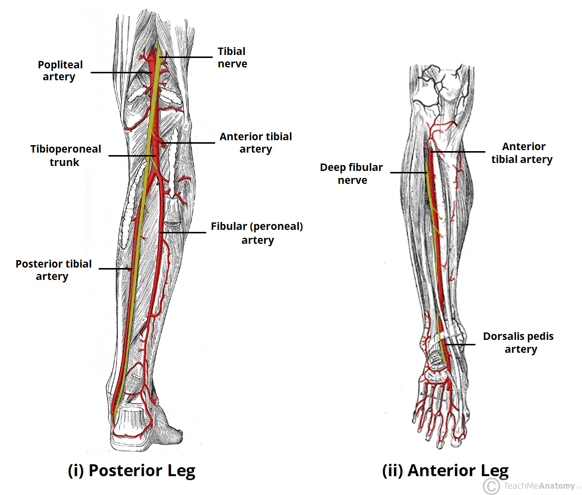
Arteries Of The Lower Limb Thigh Leg Foot Teachmeanatomy from teachmeanatomy.info Deoxygenated blood from the peripheral veins is transported back to the heart from capillaries, to venules, to veins, to the right side of the heart, and then. Bulky middle tunic contains smooth muscle and elastin 3. Learn more about the anatomy and types of blood vessels and the diseases that affect them. The popliteal artery branches in the proximal leg to ultimately form the anterior tibial, posterior tibial, and peroneal trunks. In a cadaver study, blue latex was injected into the external iliac arteries of 11 cadaveric legs and the gracilis myofasciocutaneous flap dissected. In addition, we defined the arterial anatomy of the flap that contributes to enhanced flap survival. Dorsalis pedis artery tendon of ehl m/s. To identify areas at risk during surgical interventions, we performed an anatomical study of human legs.
Arteries (in red) are the blood vessels that deliver blood to the body.
Thorough knowledge of the fascial compartments of the leg is a prerequisite of understanding the relationship between superficial and deep veins. Human anatomy and physiology ii lab practical i: Maybe you would like to learn more about one of these? Deep veins, located in the center of the leg near the leg bones, are enclosed by muscle. Common conditions that cause leg pain due to blood vessel problems are discussed below. Leg pain may occur due to problems with the arteries and/or veins in the leg. Use key choices to identify the blood vessel tunic described. Selective ink injection of the pedicle and perforating vessels also was performed in 8 legs. Spider veins are smaller versions of varicose veins, which are abnormal dilated blood vessels that often form in the legs. Deep veins , located in the center of the leg near the leg bones, are enclosed by muscle. The older term trifurcation is a misnomer because the common tibioperoneal trunk is interposed between the origin of the anterior tibial artery and the bifurcation of the other two vessels some 2 to 3 cm more distally (). Jzk 303 oled display fingertip pulse oximeter spo2 oxygen monitor for healthcare home use. Learn more about the anatomy and types of blood vessels and the diseases that affect them.
The popliteal artery branches in the proximal leg to ultimately form the anterior tibial, posterior tibial, and peroneal trunks. Reduced blood supply to the legs can cause numbness and pain that spreads into the leg while walking. Selective ink injection of the pedicle and perforating vessels also was performed in 8 legs. Deep veins, located in the center of the leg near the leg bones, are enclosed by muscle. Dorsalis pedis artery tendon of ehl m/s.
Veins Of The Foot Plantar Veins Of The Foot Labeled Canstock from cdn.xxl.thumbs.canstockphoto.com Anatomy of the lower extremity veins. The videos are done by dr. Blood vessels and nerves of the leg by dr. Blood vessels (arteries and veins)/lymphatic system. Deep veins, located in the center of the leg near the leg bones, are enclosed by muscle. The femoral artery and femoral vein — two major blood vessels — travel through the pelvic bone. Diagnosis and treatment, a panel of recognized experts comprehensively reviews the. They develop when the valves and walls of the veins are weak and cause blood to pool in the veins.
In a cadaver study, blue latex was injected into the external iliac arteries of 11 cadaveric legs and the gracilis myofasciocutaneous flap dissected.
Blood vessels in the leg may occlude, get compressed, or become inflamed. The popliteal artery branches in the proximal leg to ultimately form the anterior tibial, posterior tibial, and peroneal trunks. Deep veins , located in the center of the leg near the leg bones, are enclosed by muscle. Leg blood vessels> leg blood vessels see all 5000 products in leg blood vessels. Leg pain may occur due to problems with the arteries and/or veins in the leg. The older term trifurcation is a misnomer because the common tibioperoneal trunk is interposed between the origin of the anterior tibial artery and the bifurcation of the other two vessels some 2 to 3 cm more distally (). In addition, we defined the arterial anatomy of the flap that contributes to enhanced flap survival. A primary purpose and significant role of the vasculature is its participation in oxygenating the body. The posterior tibial artery gives off a crucial branch called the fibular/peroneal artery which mainly supplies the muscles of the leg. The videos are done by dr. Human anatomy and physiology ii lab practical i: Deep veins, located in the center of the leg near the leg bones, are enclosed by muscle. Review the major systemic veins of the body including the veins of the neck, arm, forearm, abdomen, pelvis, thigh, and leg in this.
Blood vessels 2 labeled palmar arch digital artery right femoral a right femoral v great saphenous vein left popliteal a right anterior tibial a. Vessels transport nutrients to organs/tissues and to transport wastes away from organs/tissues in the blood. Use key choices to identify the blood vessel tunic described. Tutorials and quizzes on the circulation of blood and the anatomy, structure, and physiology of blood vessels, using interactive animations and diagrams. Anatomy of blood vessels review sheet 32 261 microscopic structure of the blood vessels 1.
Blood Vessels Of Abdomen And Pelvis Anatomy Overview Kenhub from thumbor.kenhub.com Arteries and veins branch off from the femoral. Deep veins, located in the center of the leg near the leg bones, are enclosed by muscle. Tutorials and quizzes on the circulation of blood and the anatomy, structure, and physiology of blood vessels, using interactive animations and diagrams. The videos are done by dr. To identify areas at risk during surgical interventions, we performed an anatomical study of human legs. In addition, we defined the arterial anatomy of the flap that contributes to enhanced flap survival. A primary purpose and significant role of the vasculature is its participation in oxygenating the body. Blood vessels in the leg may occlude, get compressed, or become inflamed.
Blood vessels 2 labeled palmar arch digital artery right femoral a right femoral v great saphenous vein left popliteal a right anterior tibial a.
It moves through the popliteal fossa, exiting between the gastrocnemius and popliteus muscles. We did not find results for: They develop when the valves and walls of the veins are weak and cause blood to pool in the veins. These vessels transport blood to and from each leg. Anatomy_of_leg_blood_supply 1/3 anatomy of leg blood supply book anatomy of leg blood supply. This process of blood flow within your body is called circulation. Blood vessels labeled simple : Blood vessels and nerves of the leg by dr. Hip and thigh practice questions. Blood vessels 2 labeled palmar arch digital artery right femoral a right femoral v great saphenous vein left popliteal a right anterior tibial a. Jzk 303 oled display fingertip pulse oximeter spo2 oxygen monitor for healthcare home use. See all 3 sets in this study guide. Spider veins are smaller versions of varicose veins, which are abnormal dilated blood vessels that often form in the legs.
Anatomy of the lower extremity veins blood vessels labeled. Anatomy of the heart and blood vessels.


Posting Komentar
0 Komentar